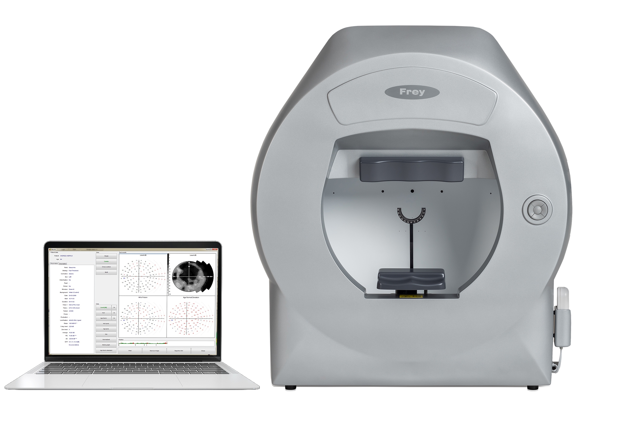
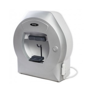
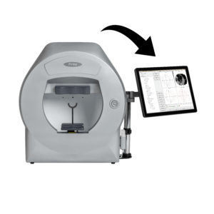
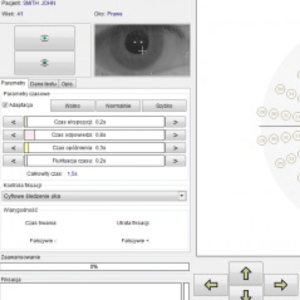
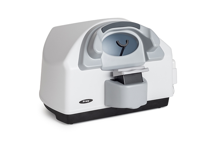
Visual field analyzer AP-250 and AP-250BY are fully functional static back LED projection automated perimeters with a full field measurement. AP-250 and AP-250BY use green color LED projection of stimulus in Goldman size III. Visual field analyzer AP-250BY additionally offers test Blue-on-yellow with a blue stimulus Goldman V size and yellow backlight in accordance with the requirements of the SWAP perimetry. The software supplied with the device offers a wide range of strategies, fields and test parameters. Control of fixation is performed automatically using the built-in camera or by controlling the position of the blind spot. Built-in data analysis include regression analysis and standardized formats for presenting and printing test results. Frey perimeter AP-250 as well as AP-250BY can be easily set up with any PC computer running the Windows operating system.
Static perimetry is the most frequently used technology for quality of visual field verification, detection of glaucoma and for monitoring glaucoma related changes in visual field. AP-250 and AP-250BY are supported by a rich library of test fields and strategies that meet and exceed user requirements and broad range of perimetry applications.
Bi-Driving test
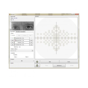
Bi-Driving is test for binocular patient examination with vision field extended to 80˚ nasally and temporally. The test fulfils requirements for driver testing. During the test, both eyes of the patient are simultaneously monitored by the digital camera.
Blue-on-Yellow
Blue-on-Yellow static perimetry is a test used for early glaucoma detection. This feature is available on AP-250BY and performed according to SWAP recommended Goldman size V stimulus in blue color presented on yellow color background.
Reduced ambient light conditions
AP-250 and AP-250BY measurement bowl is very well designed and engineered housing a combination advanced digital bowl illumination controls which reduces requirements for test room illumination conditions. For improved patient comfort, ventilation is provided by whisper-quiet concealed fans.
Simple patient positioning
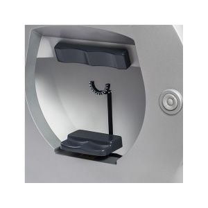
Electrically driven chinrest with movement across horizontal and vertical axis, allows for easy patient positioning. Movement of the chinrest can be controlled with push buttons located on the front of the perimeter housing or from PC with perimeter software. Anatomical head and chin support provide patient comfort during the examination.
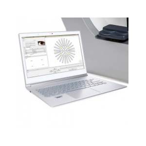
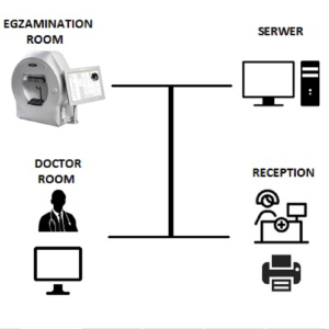
External PC
Network communication between PC and AP-250 and AP-250BY perimeters only require one USB 2.0 port, creating an effective and seamless interface with any PC running Windows. All PC network connected devices such as printers or storage servers can be used for examination test result printing or data backup and storage.
AP-250/ 250BY TECHNICAL SPECIFICATION
Device type
Automated perimeter
Measurement bowl type
Hemispherical 300mm radius with difusive surface
Maximum temporal range (degrees)
80°
Stimulus
Green-on-white
Blue-on-yellow (only AP-250BY)
Stimulus duration
0.1 – 9.9s
Stimulus size
AP-250BY – III & V
AP-250 – III
Stimulus intensity
0.03asb to 10000asb in 15 3dB or 45 1dB steps
Visual field testing distance
30cm
Background illumination
White 10asb (3.2cd/m2)
Yellow 100cd/m2 for Blue-on-Yellow (AP-250BY only) Automatic background illumination control
Threshold: Full Threshold, Fast Threshold, Smart Threshold Others: 2-Zone, 3-Zone, Quantify Defect, Constant, Binocular, Bi-Driving, Targeted Perimetry, Neurological
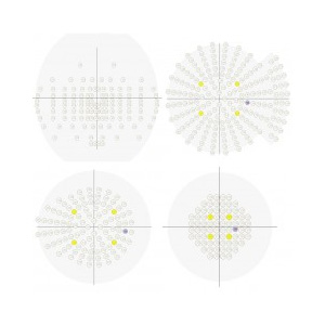
Test field library
With concentric test point positions: Macula, Central 22°, Central 30°, Full, Driving, Wide, Glaucoma, Peripheral
User defined test fields
Bi-Driving,
Industial Medicine, monocular, binocular
Correction glass diameter
38mm
Chinrest
Electrically driven in horizontal and vertical axis
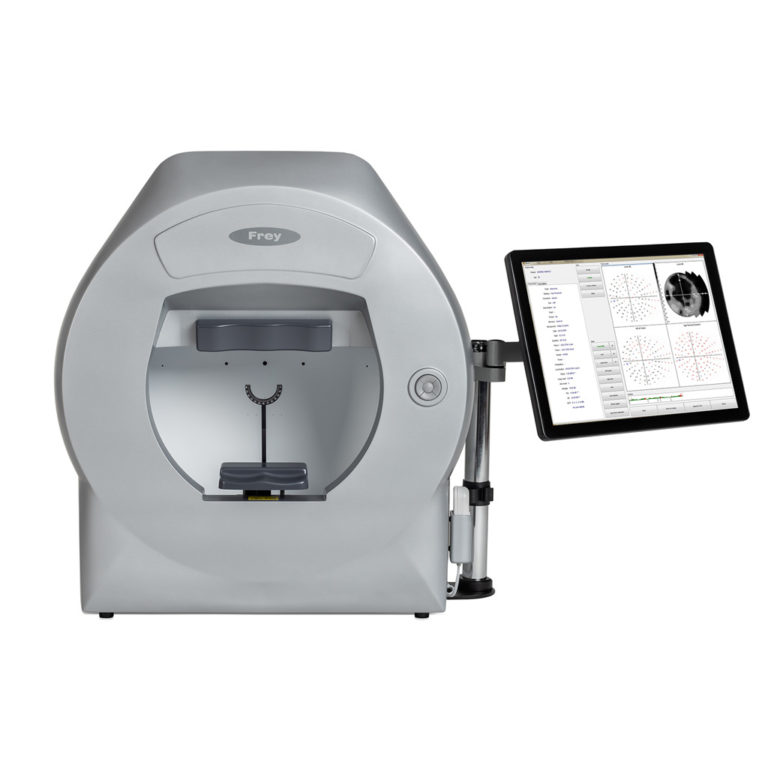
Computer
Touch screen support
Printer
External or network printer
Additional software features
Fovea threshold testing
Automatic pupil measurment
User management module
Touch screen operation
DICOM export
Network connectivity
Programming interface for EMR systems
Data import from HFA devices
Auto backup
Dimensions
Height: 637mm
Width: 566mm
Depth: 420 mm
Weight
18kg
Power requirements
Voltage 110-230VAC
Power 95W
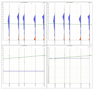
Defect progress analysis
Regression analysis module allows user to track changes in field of vision in time, using easy for interpenetration graphs. Multiple examination results of the same patient can be used for the analysis. Regression curves can be presented i absolute dB values, in relation to hill of vision or age normative values or particular global parameters like PD or AD. Analysis of the data can be limited to particular area of vision field like periphery or center, or can cover entire tested field of vision.
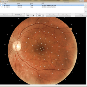
Fundus overlay
To better visualize relation of vision field defects and retinal image, Fundus overlay function allows to import fundus image and then use it as a background for displaying values recorded during the examination. Proper position of fundus image in relation to perimetry data is achieved by selecting macula and blind spot on fundus picture.
Test editor
Test field editor allows user to create their own test fields specific for particular requirements. For faster creating new test field it starts with predefined ones, like 30-2.
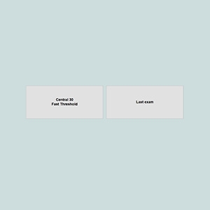
Quick access menu
For better operation efficiency quick access menu reduces number of parameters required to be entered before examination may start. User can predefined it’s test types, then operator has to select or enter patient data only. This function reduces overall test time and introduces standardization to test procedures.
HFA data import function
Migrate easily examination data to AP-250 and AP-250BY from HFA device. Import functions accepts files stored in xml and pdf formats.
Intuitive user interface
User interface of Perimetry application is designed to suport efficient work with minimum time spent on switching between options. It’s ergonomy and simplicity assures short learning time for the operator. The application is ready to be operated with use of touch screen or with mouse and keyboard depending on user preferences.
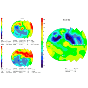
Field changes comparison
Fastest and simplest way of vision field changes is Compare function. It allows to compare results of two examinations of the same patient. Data can be presented in many available display modes like absolute values, grey scale or color maps, etc.
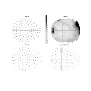
Multiple methods of examination data presentation
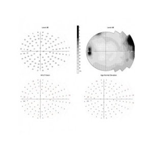
To better visualize possible vision field defects user has reach set of data presentation modes available to choose from. Result maps can be displayed as: – numeric values, pattern grade or grey scale – absolute values in dB, as relative to hill of vision, age normal related, defect probability – defect curve (Bebie graph) – normalized vales
Installation flexibility
AP-250 can be use in both small offices and large clinics. With built-in networking capabilities installation of the device is very easy and system expansion inexpensive. Perimeter can work with devices connected directly or network printers and servers. Software perimeter supplied with the device can be installed on any number of computers, allowing you to organize very comfortable working medical facility.
Data safety
Automatic backup file generator frees the operator for creating copies of examination data. Auto-backup function can start automatically with period and in location defined by the user. This function assures safety of patient records.
Data import & export
Export/Import module of Perimeter software is designed to allow the user transfer examination data between remote locations. Data may be exported in proprietary format or in compatible with EMR systems DICOM format. You can migrate easily examination data to AP-250/AP-250BY from HFA device. Import function accepts files stored in .xml and .pdf formats.
YOU MAY ALSO LIKE
How can you improve your clinical workflow?
We know the answer. Sign up to FREY newsletter. No SPAM… promise.
By entering your email address above you agree that you wish us to contact you with marketing content related to FREY. We will not share your data with anyone else or use it for any other purpose. Read our Privacy Policy notice for more information. You may have your details removed from our database at any time by contacting contact@freymedical.eu.