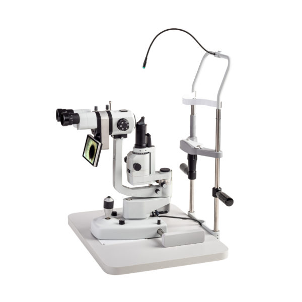
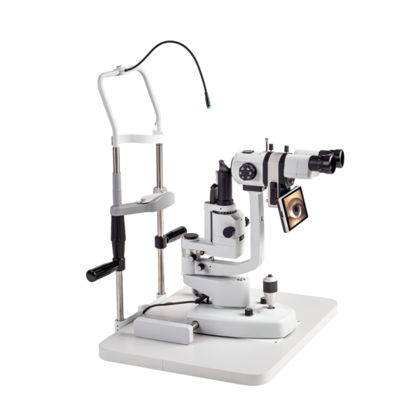
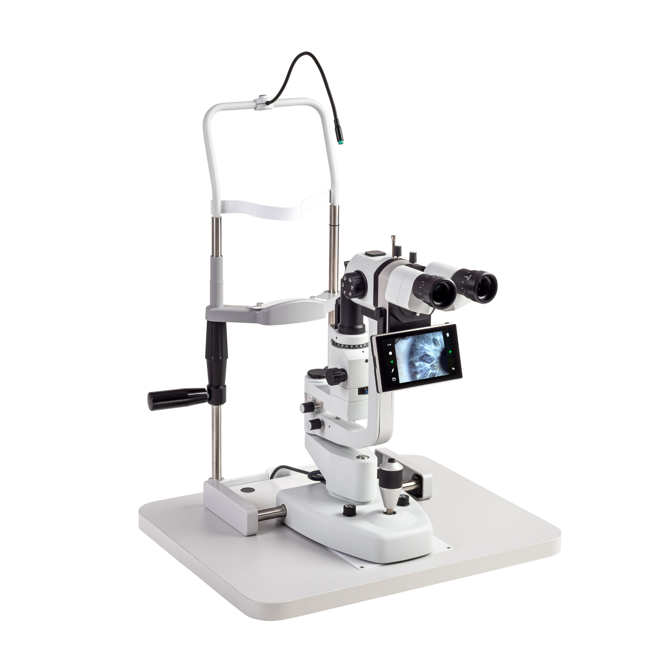
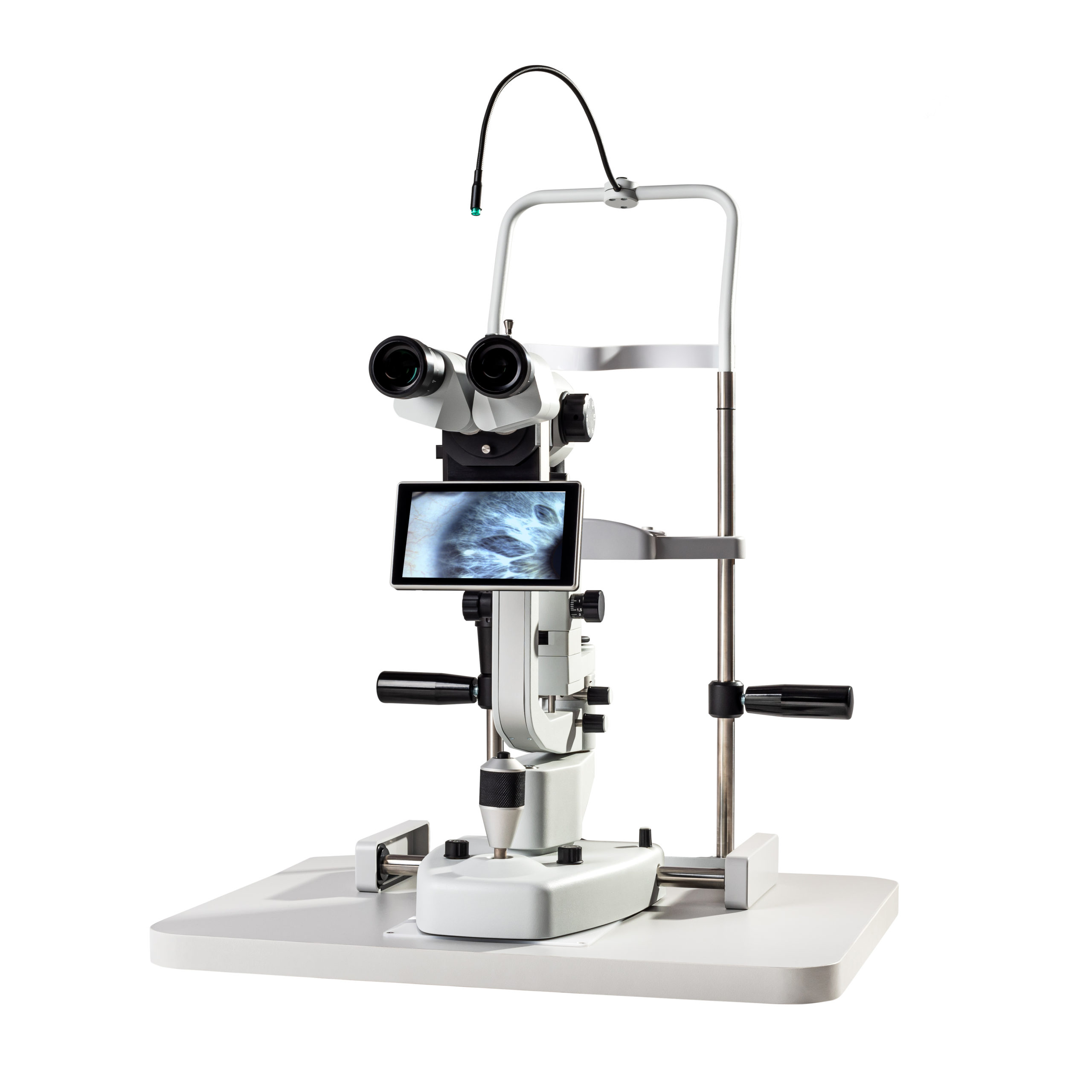
Digital Slit Lamps & Imaging Systems

SL-100
SL-100 & SLI-200
SL-100 & SLI-200 4K
SL-110
SL-110 & SLI-200
SL-110 & SLI-200 4K

FREY DIGITAL SLIT LAMPS

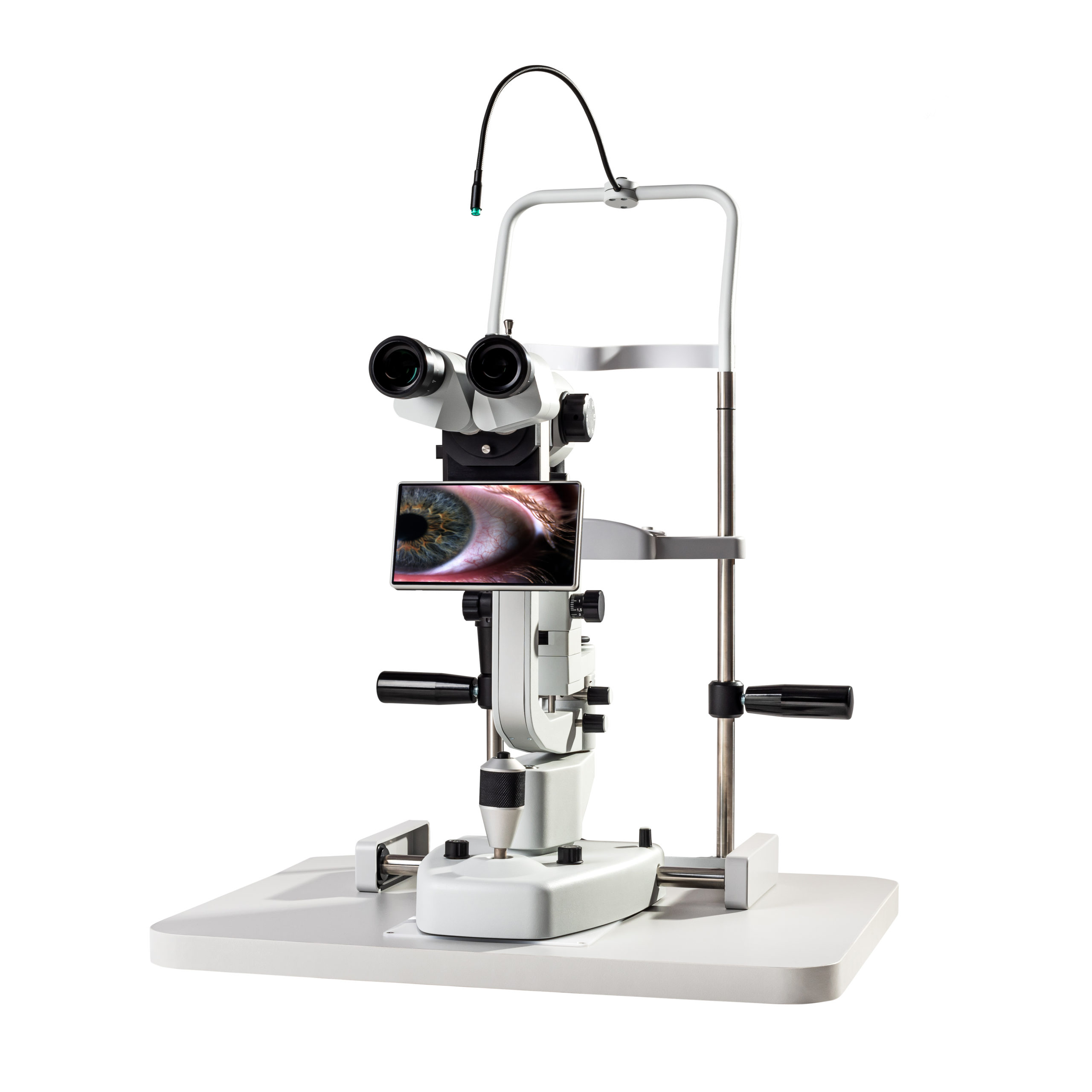
FREY digital slit lamps provide par excellence clinician experience by embracing the latest in diagnostics combined with superb engineering and state-of-the-art technology. Images and videos are now conveniently accessed without the need to acquire and install additional peripherals

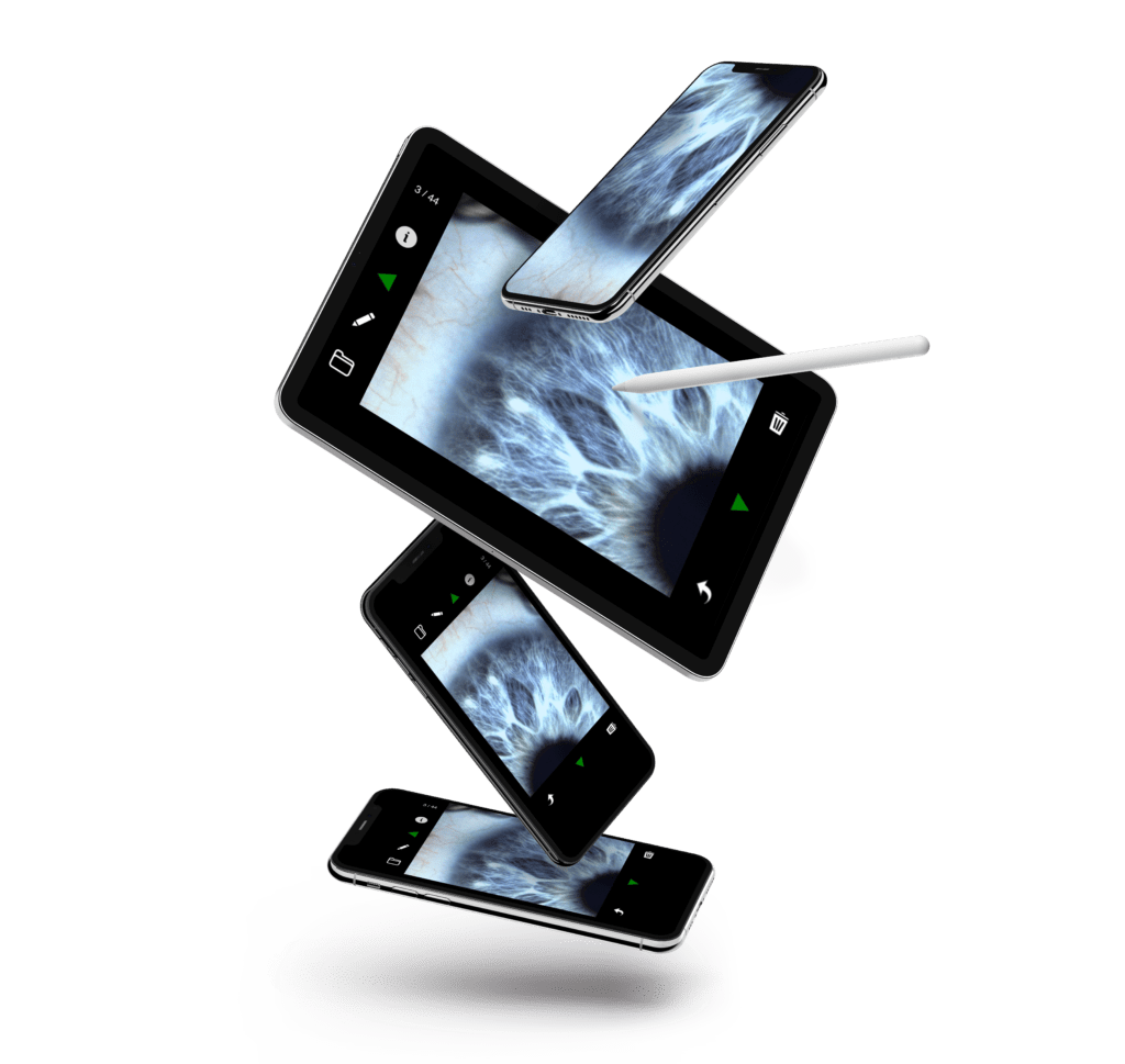
EYEMAGER – dedicated software application, can send, clinical results directly to any networked device – smartphone, laptop, tablet or computer, safely and securely, anytime, anywhere FREY devices are designed to enable clinician workflow to be more effective and efficient across multiple workplaces.  FREY digital slit lamps deliver an optimized experience that is outstanding.
Optimize clinical space. Improve workflow.

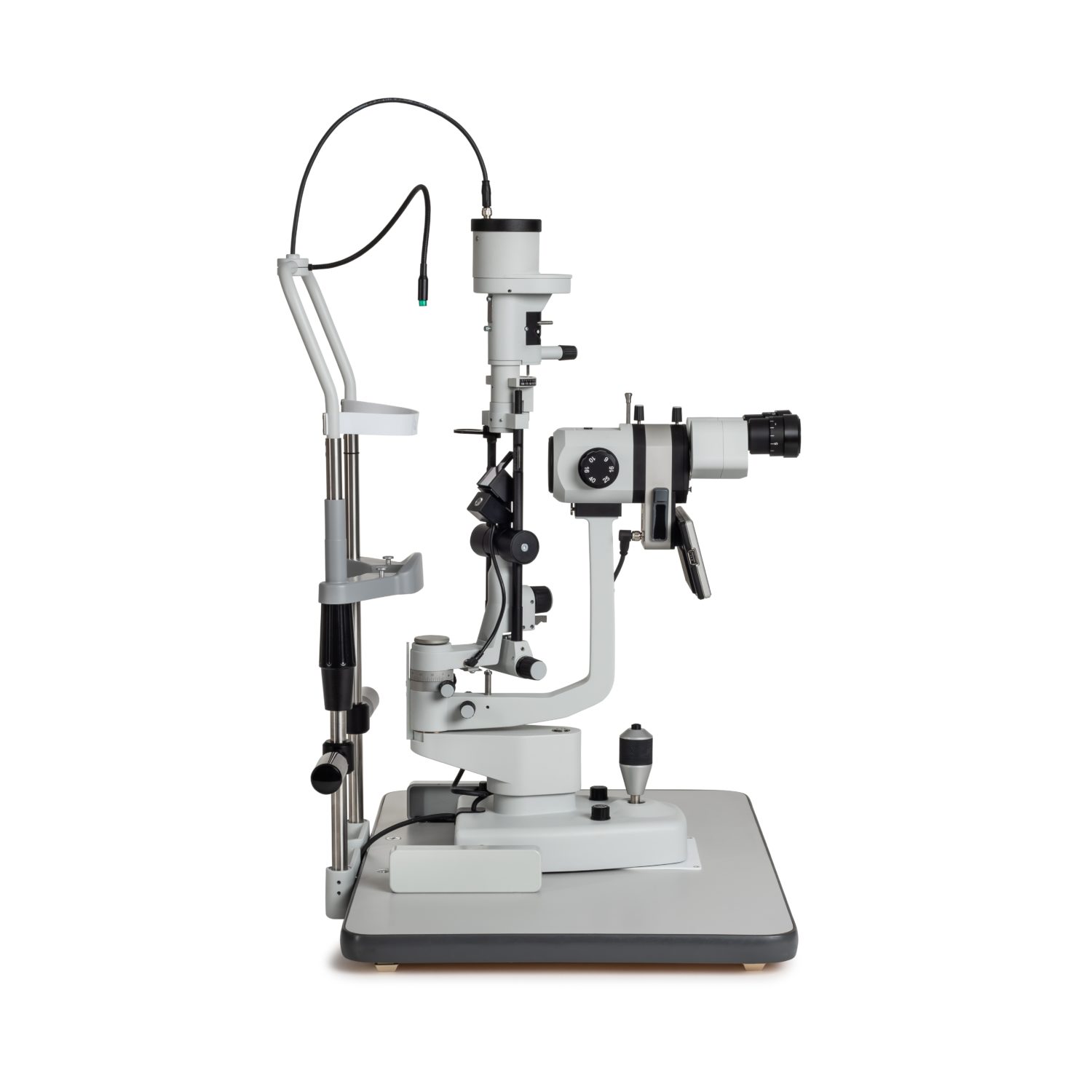
FREY slit lamps are elegantly designed for digital imaging and visualization and provide an amazing user experience in comfort. Intelligent design control functions and components are fully integrated, with cabling subtly concealed inside the slit lamp body. Capture high resolution images and videos with integrated or dedicated imaging systems. FREY precision LED digital slit lamps exhibit a small footprint which optimizes clinical space, and users can improve workflow more effectively and efficiently. A world class fully optioned digital slit lamp with cutting edge design and innovation designed for the modern thinking eye care professional.

Digital slit lamps are essential for ophthalmic practice.
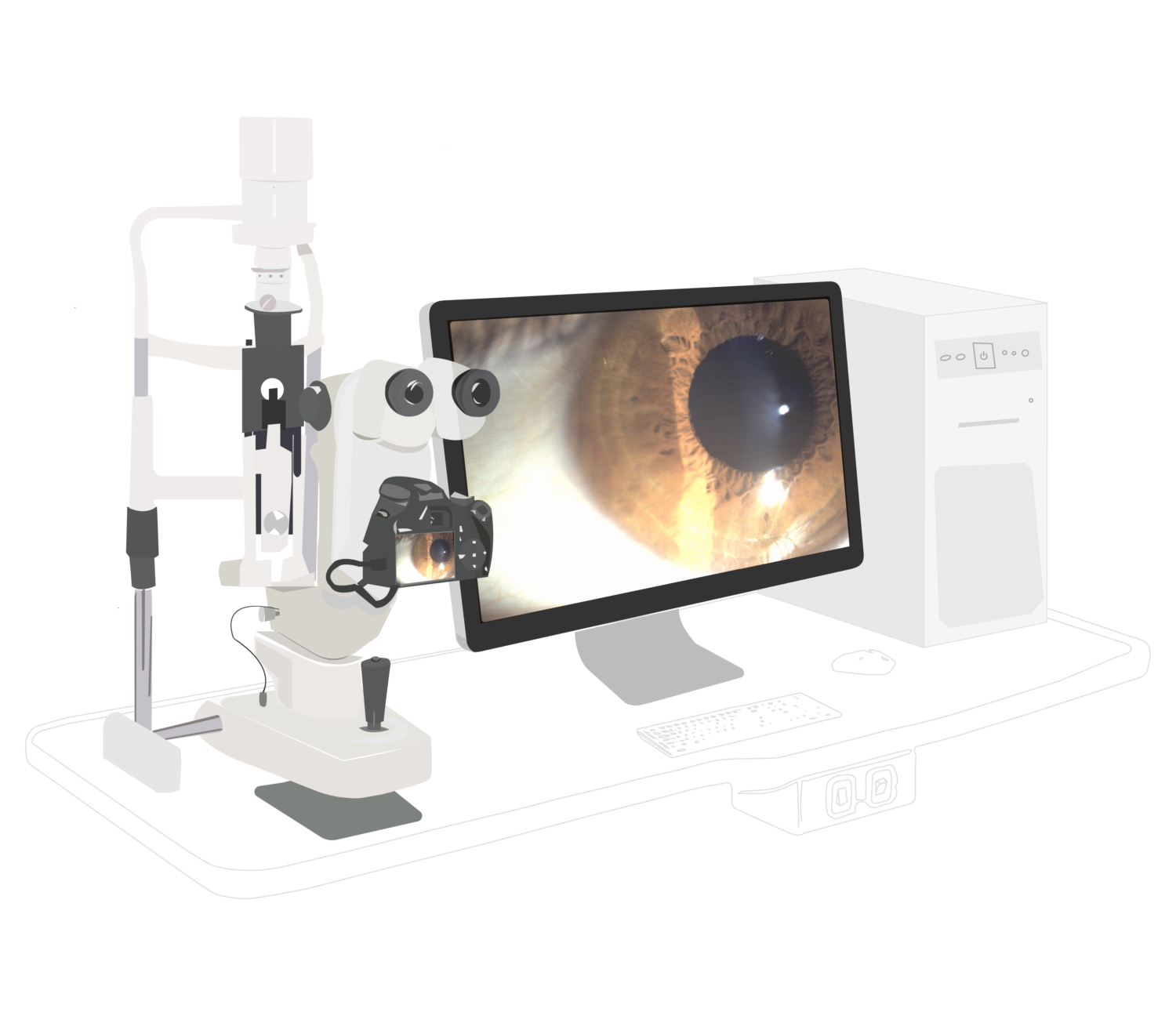
Conventional Digital Slit Lamp System
Save the workspace with FREY tools for ophthalmologists.
Space-saving FREY Digital Slit lamp system

Stunning optical quality. Visualize and diagnose more.

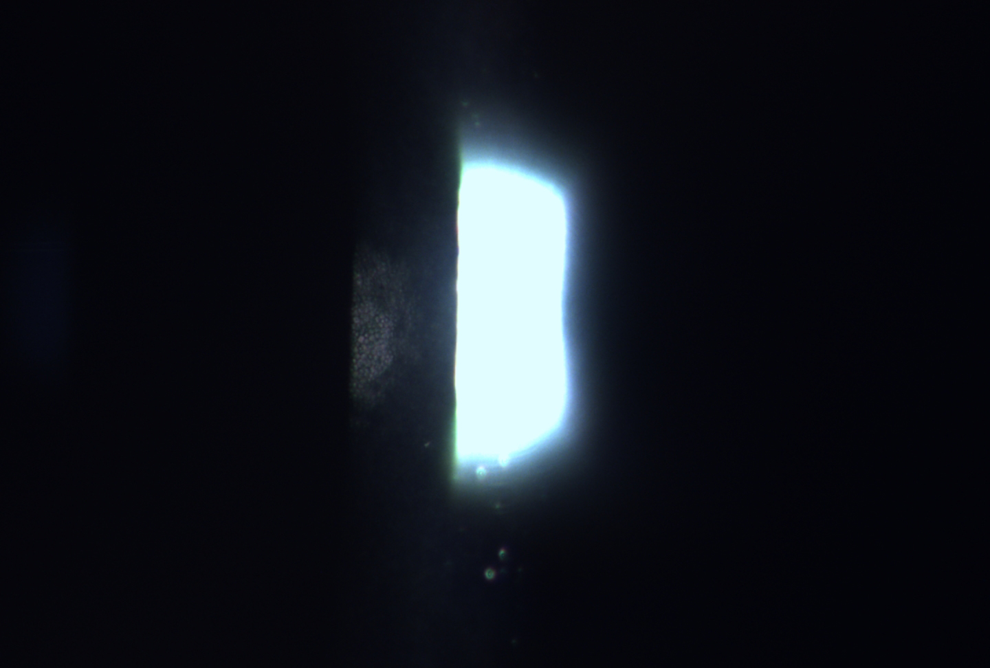
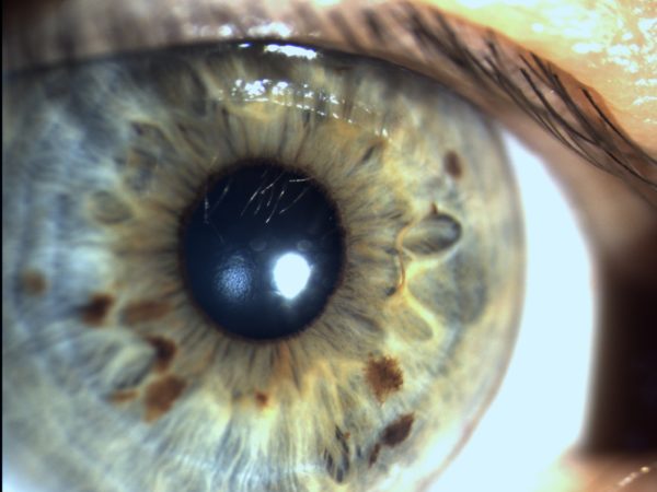
Corneal endothelium consists of a single layer of tiny hexagonal cells. Slit lamps are diagnostic instruments widely used by ophthalmologists and optometrists to observe anterior and posterior segments. Specular reflection illumination technic is used to observe cornea posterior surface (endothelium). Stunning optical quality of FREY digital slit lamps feature advanced high-sensitivity and high-resolution camera sensors to deliver superb image quality, capturing the finest of detail and visualization of individual endothelium cells.

Endothelial cells captured with FREY Digital Slit Lamp
Endothelial cells captured with FREY Digital Slit Lamp (click to enlarge photo).

Bring hidden details to (infrared) light.

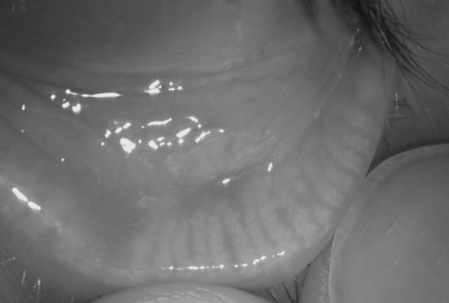
A leading cause of dry eye, Meibomian Gland Dysfunction (MGD) affects the essential oil layer of the eye’s tear film, causing tears to evaporate too quickly. For optimal treatment MGD must be clinically observed and diagnosed with certainty and clarity, allowing eye care professionals to prescribe the most appropriate treatment. A diagnostic method of MGD is examination
with a digital slit lamp equipped with infrared light (IR light source). Infrared light is not visible to human eyes. To observe the image, IR sensitive camera and a display is clinically essential. FREY digital slit lamps are equipped with optional IR illumination source to visualize MGD with superb clarity, without causing patient discomfort. FREY hi-res IR sensitive camera quickly acquires grey scale images of meibomian gland structures for early diagnosis of MGD.

Meibomian Glands captured with FREY Digital Slit Lamp.

Proprietary software. EYEMAGER.​

Eyemager™ is a proprietary imaging software designed exclusively for FREY digital slit lamps. Capture high-quality images and videos directly during examination and extract the finest details of ocular structures – including frame-by-frame video analysis for precise clinical evaluation. Manage patient records and build professional examination reports – all in one place, without additional peripherals.

Available as a built-in application on the SLI-200 and as a dedicated PC application for SLI-200 4K.


Eyemager™ features:

  • Real-time image and video preview during examination
  • Photo and video capture
  • Patient records and examination history management
  • Anonymous examination mode
  • Professional report generation (PDF and DICOM formats)

Integrated diagnostic platform. EyeExpert

EyeExpert is an optional add-on for FREY Digital Slit Lamps that connects your device to a clinic-wide data management system. It eliminates manual data entry, reduces transcription errors, and gives instant access to complete examination results and images from any authorised workstation in your clinic. Everything runs on your local network.

EyeExpert DataHub

DataHub is the server application at the heart of the EyeExpert ecosystem. It runs on a single clinic server and manages everything: patient records, image archives, user accounts, and connected devices. Reception staff schedule visits directly from a web browser – appointments flow automatically to the slit lamp, with no manual data entry at the device. The moment an examination ends, results and images are archived and ready to review. No USB drives, no manual export, no waiting.

  • Visit scheduling and worklist management — schedule visits from any browser on the network; appointments appear automatically on the slit lamp
  • Automatic result archiving — images and videos land in the archive the moment the examination ends
  • Central patient database — one record for every patient, shared across all devices in the clinic
  • Role-based user accounts — separate permissions for standard users and administrators
  • Continuous backup — automatic backup to external storage for data safet

EyeExpert Viewer

EyeExpert Viewer is a Windows desktop application installed on physician workstations across the clinic. It connects to DataHub over the network and gives immediate access to the full examination history of every patient –  slit lamp images, video recordings, and reports – right at the physician’s desk.

  • Image and video review — browse captured images, frame series, and video recordings with zoom, pan, and frame-by-frame playback
  • Both eyes, side by side — right and left eye results displayed simultaneously for direct comparison
  • Scrollable visit history — all examinations listed chronologically; scroll through the patient’s full history at a glance
  • Area annotations on images — mark specific regions with circles or rectangles and add clinical notes directly on the image
  • Physician annotations — add global notes to individual results or flag them as unreliable
  • Printable PDF reports — save or print reports directly from the Reports panel
  • Works on any clinic workstation — install once per Windows PC; connects to DataHub automaticall
Important: EyeExpert Viewer requires a running EyeExpert DataHub instance. DataHub acts as the authentication server, data store, and connection broker for all Viewer installations. Install DataHub first — enter its server address in Viewer once to connect.

BROCHURES

VIDEOS

This website is intended for healthcare professionals and authorised distributors only. © 2026 Frey S.J. All rights reserved.